


A microscopic tool with macro potential
UTSW provides scientists with access to cutting-edge 3D electron microscopy that permits the deepest looks inside cells to advance biomedical research

In the 1930s, an incredible innovation forever changed scientific research: the electron microscope, which gathers images with beams of electrons rather than optical microscopes’ visible light. Because electrons’ wavelengths are significantly shorter than those of visible light, electron microscopes provided unprecedented resolution, allowing researchers to peer inside biological structures at the molecular level.
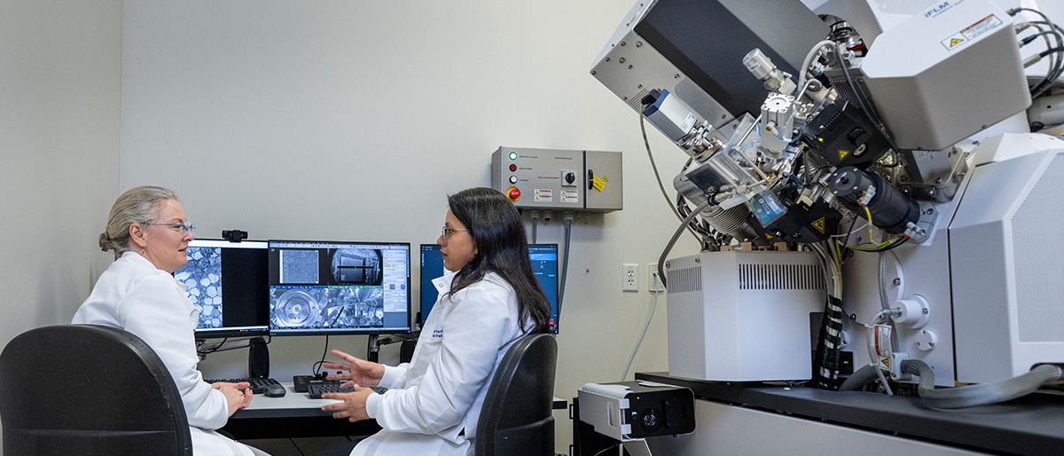
Although electron microscopy has facilitated countless scientific discoveries – an achievement that won its discoverers the Nobel Prize in Physics in 1986 – it comes with a significant flaw: The images it produces are two-dimensional while biology is three-dimensional, explained Natalia Gunko, Ph.D., Associate Professor of Cell Biology and Director of the Electron Microscopy Core Facility at UT Southwestern.
“The way life exists is not the way we have traditionally seen it in electron microscopy, limiting the advances we can make,” she said.
But now electron microscopy technologies developed over the past three decades, known as volume electron microscopy (vEM) and correlative light and electron microscopy (CLEM), are helping scientists see the biological world in a whole new light.
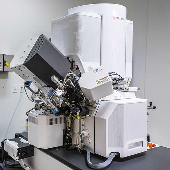
In 2025, UTSW acquired a state-of-the-art Thermo Scientific Hydra Bio Plasma FIB-SEM (Hydra) that supports both vEM and CLEM workflows – the first instrument of its kind installed in Texas. About the size of a large refrigerator, this machine contains a vacuum chamber that houses the sample stage, along with two precisely aligned beams: an electron beam for imaging and a focused ion beam used to mill away ultrathin layers of the sample during three-dimensional reconstruction. Having this groundbreaking, dual-beam equipment on campus is revolutionizing research, its users say, providing a crucial tool to make discoveries that were impossible with previous imaging approaches.
Seeing deeper

“Much of cell biology research is simply asking what the inside of a cell looks like. But traditional electron microscopy images of cell interiors often look like a bunch of squiggly spaghetti,” said William Prinz, Ph.D., Chair and Professor of Cell Biology.
Cells are three-dimensional objects jam-packed with various structures, he explained – and even by microscopic standards, their interiors are relatively large. Thus, finding the structure a scientist is interested in, and identifying the biological processes affecting it, can be a challenge with the electron microscopy approaches researchers have used for decades. More and more scientists in cell biology and other life sciences are looking to vEM and CLEM to solve this problem.
In vEM, a relatively large specimen – such as a chunk of tissue containing many cells, or even a single cell, which is a bulky object by microscopy standards – is imaged as a series of nanoscale layers (about 1,000 times thinner than the width of a human hair). Depending on the method, layers may be physically sectioned or, as with the Hydra, milled away one at a time with an ion beam while an electron beam images each newly exposed surface. Computer algorithms then reconstruct these pictures into a three-dimensional image, allowing scientists to see the biological context of what they’re studying. For example, it might show which other cellular structures surround a structure of interest, or how various cells in a tissue connect or interact.

CLEM links the ability of light microscopy to home in on chemical tags, such as fluorescent dyes, with the high resolution of electron microscopy. Chemical tags allow researchers to find a specific molecule, protein, or structure within a cell using light microscopy – the biological equivalent of finding a needle in a haystack – and image just that region with electron microscopy.
Both vEM and CLEM provide far more detailed and contextual information than light microscopy or traditional electron microscopy can deliver alone, said Deborah Carlson, Ph.D., Assistant Dean for Research Support Facilities and Associate Professor of Surgery and Pediatrics. “In layman’s terms, the difference between these new approaches and earlier electron microscopy techniques is like going from driving a Kia to a BMW,” she explained. “With vEM and CLEM, we can collect a lot more data and do it much quicker.”
Rare in-demand technology

Although both vEM and CLEM are increasingly in demand, relatively few microscopes that perform these techniques are available in the U.S. The expertise to operate them is similarly rare, explained Dr. Gunko. That’s why UTSW hired her to run the Electron Microscopy Core in 2023 and acquire a new microscope with vEM and CLEM capabilities.
A neuroscientist who has published extensively in her field, Dr. Gunko is also an expert on electron microscopy – knowledge she gained while conducting her own research during her Ph.D. program at the University of Groningen in the Netherlands and in two postdoctoral fellowships at the University of California, San Diego and the Scripps Research Institute. The Agency for Science, Technology and Research (A*STAR) in Singapore hired her to manage its Electron Microscopy Unit in 2012. Three years later, she got the opportunity to head up the Electron Microscopy Core Facility at KU Leuven, Belgium’s oldest research university, where she stayed until joining UTSW’s faculty.
“Acquiring our newest microscope began not with buying the hardware but with the good fortune to recruit Dr. Gunko,” said Joan Conaway, Ph.D., Vice Provost and Dean of Basic Research. “It’s one of many examples of how UTSW is committed to ensuring that investigators on campus have access to the most advanced technology that enables them to address questions that are important.”
A campuswide survey performed before Dr. Gunko arrived showed that many Principal Investigators at UTSW would consider a microscope with vEM/CLEM capabilities on campus a game changer for their research. Several were already using these technologies with the help of collaborators who had these microscopes on their own campuses. But having a microscope here would dramatically improve access to vEM and CLEM, accelerating research through proximity to the equipment and Dr. Gunko’s team. Working hands-on with researchers, Electron Microscopy Core Facility staff provide access to the microscopes and help investigators plan experiments, prep samples, gather data, and even write manuscripts.
“Working with collaborators at other institutions, it took us a year and a half to acquire the vEM images we needed for our studies,” said Lu Sun, Ph.D., Assistant Professor of Molecular Biology. “But with our new microscope on campus, it takes about a week.”
From glia to cilia
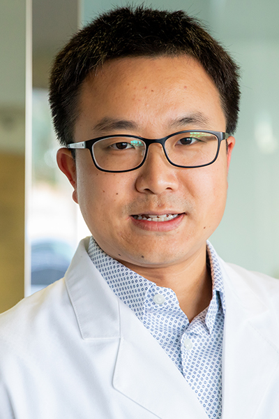
Dr. Sun’s work focuses on interactions between neurons and glia, a diverse group of cells that support and engage with neurons. A subtype of glia, known as oligodendrocytes, is responsible for producing myelin, a fatty material that coats neurons’ long extensions and functions as an insulator to help speed neurons’ electrical signaling.
Several years ago, research in the Sun Lab, led by postdoctoral researcher Daniela Barbosa, Ph.D., found that removing a specific protein called transcription factor EB caused this insulation to develop numerous defects known as myelin whorls. Similar defects are present in the myelin of patients with demyelinating diseases, such as multiple sclerosis.
Seeking to learn more about myelin whorls, the researchers needed to take a closer look – but traditional electron microscopy wouldn’t be enough, said Dr. Barbosa. Although this technology could reveal their presence on neuron extensions, its two-dimensional nature couldn’t shed light on the whorls’ size, contents, or even whether they were attached to the oligodendrocytes or free-floating.
To answer these questions, the researchers worked with collaborators at the University of California, San Diego to perform vEM. Their findings revealed that newly formed oligodendrocytes utilize this transcription factor and another downstream mechanism known as autophagy to facilitate myelin whorl removal, thereby establishing myelin integrity during neural development.
The Sun Lab plans to continue studying myelin whorls using UTSW’s Hydra.
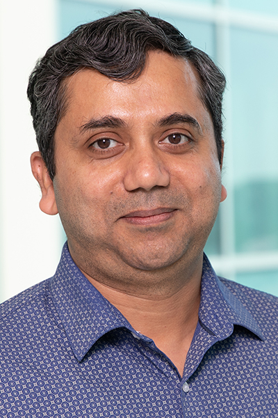
Saikat Mukhopadhyay, M.D., Ph.D., Associate Professor of Cell Biology, has used a combination of vEM and CLEM in his studies on structures called primary cilia. These organelles – which play a variety of roles, such as sensors of chemical or mechanical stimuli and hubs for molecular signaling – are found on or within nearly all cell types in the body.
He and his colleagues in the Mukhopadhyay Lab are most interested in primary cilia in the cerebellum, a brain region in the back of the head. As the cerebellum grows and develops after birth, the major neuron progenitors in the cerebellum are peppered with primary cilia; however, these structures gradually disappear as neurons form and mature. Researchers in the Mukhopadhyay Lab and others have found that primary cilia reappear in the cerebellum in a form of childhood brain cancer called medulloblastoma, where they seem critical to tumor cell survival and growth.
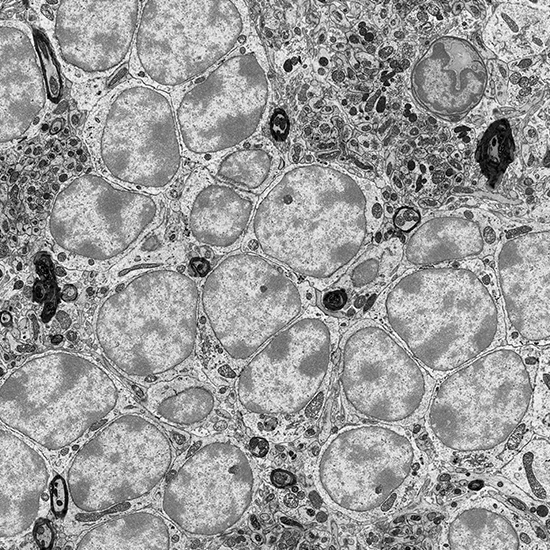
To better characterize these structures in both healthy and disease states, he and his colleagues at UTSW and collaborating institutions are using vEM and CLEM together to focus in on primary cilia in cerebellar neurons and identify other cellular structures and proteins in and around them – work that could potentially turn up therapeutic targets for medulloblastoma.
“Some of our cerebellum samples were used to calibrate the new microscope,” Dr. Mukhopadhyay said. “I can’t wait to use this machine to further our research.”
Advancing disease research
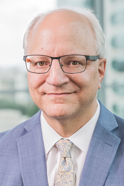
William Dauer, M.D., Director of the Peter O’Donnell Jr. Brain Institute and a Professor of Neurology and Neuroscience, also uses vEM and CLEM in his work. The Dauer Lab studies DYT1 dystonia, a developmental disease that causes involuntary muscle contractions that begin in childhood and worsen with age. Years ago, Dr. Dauer and his colleagues discovered that the mutation responsible for causing DYT1 dystonia impairs function of a protein called torsinA. Light microscopy identified abnormalities of the membrane surrounding the cell nucleus in torsinA mutant neurons and central nervous system tissue. Standard electron microscopy revealed defects in portions of this membrane associated with nuclear pores, structures that allow molecular interchanges between the nucleus and the rest of the cell. However, the exact morphology and cause of these abnormalities, and the role of defective torsinA in creating them, has been unclear.
To explore these questions – critical for developing future treatments – researchers in the Dauer Lab have used vEM, enabling them to define a role for torsinA in nuclear pore biogenesis. Dr. Dauer and his colleagues are planning to perform CLEM studies to pinpoint the location of torsinA during nuclear pore biogenesis as well as that of recently discovered proteins involved in this process.
Both technologies will play valuable roles in many other facets of neuroscience, Dr. Dauer said.
“Developing a new understanding of connectivity between cells, explaining how neural circuits work, and delving into subcellular architecture are all things we can do here, now that we have this equipment on campus,” he explained.
CLEM and vEM have the potential to accelerate advances in biomedicine, Dr. Gunko added. Researchers across campus have kept both the Hydra and the Electron Microscopy Core staff busy since the microscope’s launch in April 2025. Its findings are currently supporting more than 10 research projects across UTSW.
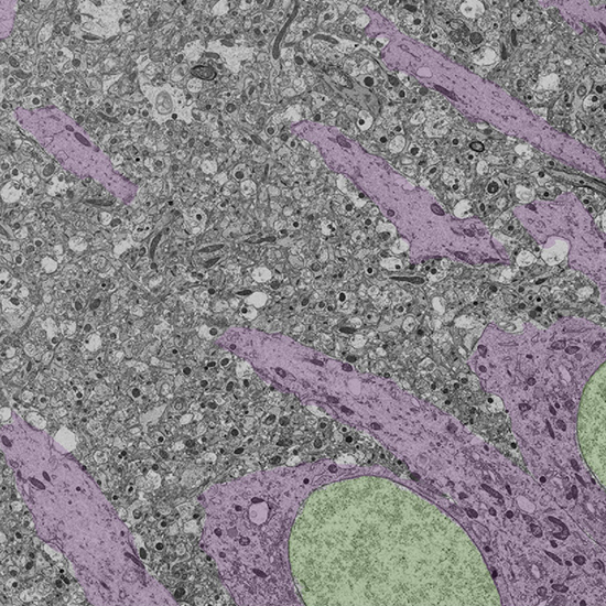
Recently, the Electron Microscopy Core received a grant from the National Institutes of Health to purchase another microscope with different capabilities: The JEOL JEM-120i 120 kV Transmission Electron Microscope, expected to be delivered to UTSW in January 2027, will image biological and other soft materials at resolutions approaching the atomic scale by transmitting electrons through ultrathin specimens. Dr. Gunko expects these technologies will further improve over time, with abilities to analyze even larger volumes of tissue and incorporate artificial intelligence to better identify items of interest on the horizon.
“To have this equipment available on campus is an incredible gift for our researchers, literally,” said Dr. Gunko, adding that she purchased the Hydra during the holiday season, on New Year’s Eve 2024. “With this microscope, we have the potential to do so much more than we ever have before.”
Endowed Titles
Dr. Conaway holds the Cecil H. Green Distinguished Chair in Cellular and Molecular Biology.
Dr. Dauer holds the Lois C.A. and Darwin E. Smith Distinguished Chair in Neurological Mobility Research.
Dr. Mukhopadhyay is a W. W. Caruth, Jr. Scholar in Biomedical Research.
Dr. Prinz holds the Virginia and Edward Linthicum Distinguished University Chair in Biomedical Science.
Dr. Sun is a Southwestern Medical Foundation Scholar in Biomedical Research.