
Microscope overview
The QLMC provides access to a wide range of advanced imaging modalities, with particular expertise in confocal microscopy, super-resolution imaging, fluorescence lifetime imaging microscopy (FLIM), and holotomography. To maximize resources and avoid duplication of services, the QLMC works closely with complementary core facilities across the UTSW campus. If you are unable to identify a suitable imaging platform, or are unsure which technology would be most appropriate, don’t hesitate to contact us – we’re happy to discuss experimental requirements and help identify the most suitable imaging solution, whether within the QLMC or through one of our collaborating core facilities.
Explore the imaging systems offered by the QLMC. Select Technical details for additional specifications.Color legend:● Super-resolution● Confocal● Widefield fluorescence● Holotomography● Stereo / dissecting

Abberior Facility Line STED
Super-resolution · Inverted
3D STED super-resolution microscope with adaptive illumination and adaptive optics for imaging deeper specimens.
Manufacturer: Abberior Instruments
Location: ND5.760
Internal rate: $25/hour
$25 autonomous; $65 assisted
Key capabilities: Confocal; 2D STED; 3D STED; Adaptive illumination; Adaptive optics; RESCue; Tiling
Technical details ▾
| Imaging modalities | Confocal; 2D STED; 3D STED |
|---|---|
| Super-resolution | STED |
| Specimen types | Live cells; Fixed cells; Tissue. |
| Sample formats | Slides; 35 mm dishes |
| Excitation | 405; 485; 561; 640; depletion: 595, 775 |
| Environmental control | Live-cell: Yes; temperature: Yes; CO₂: Yes; humidity: Yes |
| Special capabilities | Adaptive illumination; Adaptive optics; RESCue; DyMIN; MINFIELD; Automated z-stacks; FRET; Tiling |
| Objectives | 60x/1.42 oil; 60x/1.20 water; 60x/1.30 silicon |
| Recommended applications | 2D STED; 3D STED; Organoids super-resolution. |
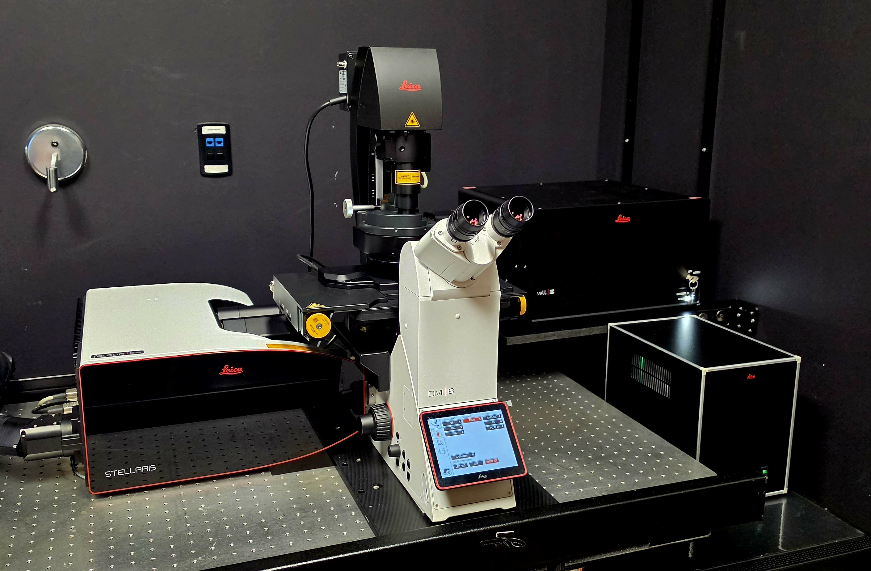
Leica Stellaris Falcon 8
Confocal · Inverted
Super-resolution
Inverted laser-scanning confocal microscope with integrated Falcon 8 FLIM and TauSense technology.
Manufacturer: Leica Microsystems
Location: NL5.120R
Internal rate: $40/hour
Half price outside internal peak hours
Key capabilities: Laser-scanning confocal; FLIM; FCS; TauSense; Spectral imaging; FLIM-FRET; FRAP
Technical details ▾
| Imaging modalities | Laser-scanning confocal; FCS; FLIM; Spectral imaging |
|---|---|
| Super-resolution | LIGHTNING |
| Specimen types | Immunofluorescent cell cultures; Tissue sections, Organoids; Live cells; Fixed cells |
| Sample formats | Slides; 35 mm dishes; Multi-well chambers; Multi-well plates |
| Excitation | 405 plus 440–790 (tunable White Light Laser) |
| Detectors | 5 detectors: 3 HyD S + 1 HyD X + 1 HyD R; detection range 400–850 nm |
| Environmental control | Live-cell: Yes; temperature: Yes; CO₂: Yes; humidity: Yes |
| Special capabilities | FLIM; TauSense; Spectral imaging; Spectral unmixing; FLIM-FRET; FRAP; FRET; FCS; Photoablation; Photoactivation; Uncaging; infrared imaging; Resonant scanning; Simultaneous multi-channel acquisition; Fast z-stacks; Tiling; Focus stabilization |
| Objectives | 10x\0.4 air; 20x\0.75 air; 40x\0.95 air; 40x\1.3 oil; 63x\1.4 oil |
| Recommended applications | Live-cell imaging; Fixed-cell imaging; Tissue; Fluorescence lifetime imaging; Spectral imaging |
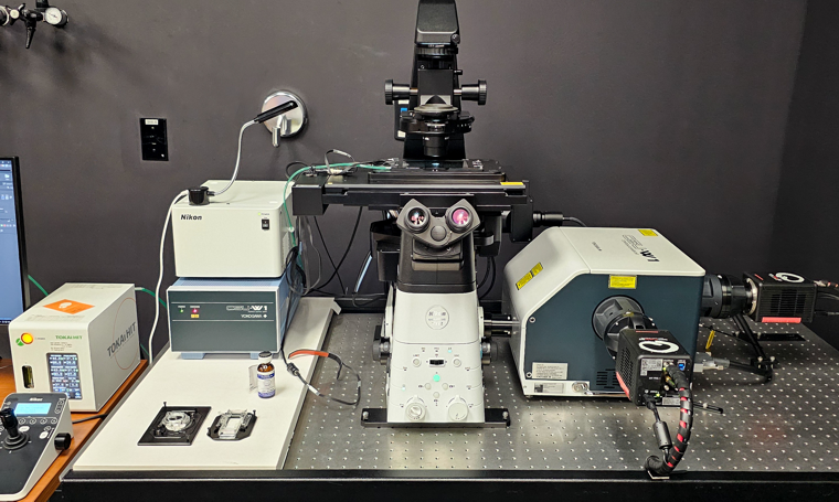
Nikon CSU-W1 Dual Camera
Confocal · Inverted
Fast inverted spinning-disk confocal microscope with two sCMOS cameras for simultaneous two-channel imaging.
Manufacturer: Nikon
Location: K1.228
Internal rate: $40/hour
Half price outside internal peak hours
Key capabilities: Spinning-disk confocal; Widefield fluorescence; Simultaneous two-channel acquisition; Fast z-stacks; Tiling; Focus stabilization
Technical details ▾
| Imaging modalities | Spinning-disk confocal; Widefield fluorescence; Brightfield |
|---|---|
| Specimen types | Immunofluorescent cell cultures; Tissue sections, Organoids; Live cells; Fixed cells |
| Sample formats | Slides; 35 mm dishes; Multi-well chambers; Multi-well plates |
| Excitation | 405; 445; 488; 514; 561; 594; 640 |
| Detectors | sCMOS |
| Cameras | 2 × Hamamatsu ORCA-Fusion BT |
| Transmitted light | Phase contrast; DIC; Brightfield |
| Environmental control | Live-cell: Yes; temperature: Yes; CO₂: Yes; humidity: Yes |
| Special capabilities | Simultaneous two-channel acquisition; Fast z-stacks; Automated z-stacks; FRET; Tiling; Focus stabilization; Up to 100 frames/s |
| Objectives | 4x\0.2 air; 10x\0.45 air; 20x\0.8 air; 40x\1.3 oil; 60x1.42 oil; 100x\1.45 oil |
| Recommended applications | Fast live-cell imaging; Rapid cellular dynamics; Simultaneous two-color imaging; Fixed-cell imaging; fast multicolor z-stacks |
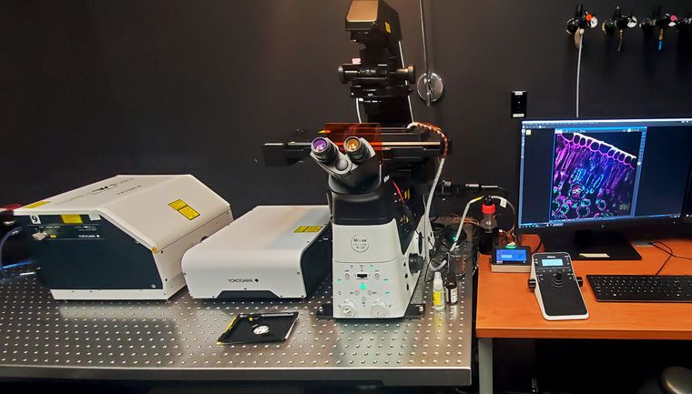
Nikon CSU-W1 SoRa
Confocal · Inverted
Super-resolution
Fast spinning-disk confocal microscope with SoRa super-resolution and a dedicated FRAP module.
Manufacturer: Nikon
Location: NL5.120R
Internal rate: $40/hour
Half price outside internal peak hours
Key capabilities: Spinning-disk confocal; Widefield fluorescence; FRAP; SoRa; Photoablation; Photoactivation
Technical details ▾
| Imaging modalities | Spinning-disk confocal; Widefield fluorescence; Brightfield; Polarized light |
|---|---|
| Super-resolution | SoRa |
| Specimen types | Immunofluorescent cell cultures; Tissue sections, Organoids; Live cells; Fixed cells |
| Sample formats | Slides; 35 mm dishes; Multi-well chambers; Multi-well plates |
| Excitation | 405; 445; 488; 514; 561; 640; FRAP: 405, 473, 561 |
| Detectors | sCMOS |
| Cameras | Hamamatsu ORCA-Fusion |
| Transmitted light | Phase contrast; DIC; Brightfield; Polarized light |
| Environmental control | Live-cell: Yes; temperature: Yes; CO₂: Yes; humidity: Yes |
| Special capabilities | SoRa; FRAP; FRET; Photoablation; Photoactivation; Uncaging; Fast z-stacks; Automated z-stacks; Tiling; Focus stabilization; Up to 100 frames/s |
| Objectives | 4x\0.13 air; 10x\0.3 air; 20x\0.75 air; 40x\1.3 oil; 60x\1.4 oil; 100x1.45 oil |
| Recommended applications | Fast live-cell imaging; Rapid cellular dynamics; Fast FRAP recoveries; Fixed-cell imaging; fast multicolor z-stacks |
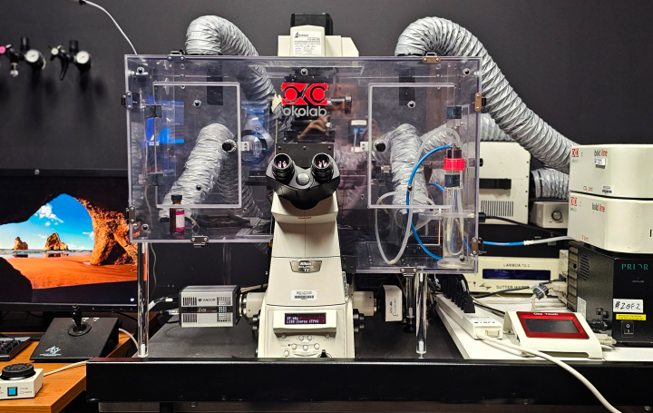
Nikon Ti Widefield Epi-scope
Epi fluorescence · Inverted
Inverted widefield microscope with monochrome fluorescence and true-color cameras for fluorescence, histology, and polarized-light imaging.
Manufacturer: Nikon
Location: NL5.120R
Internal rate: $25/hour
Standard internal rate
Key capabilities: Widefield fluorescence; Brightfield; Histology imaging; Polarized light; Focus stabilization
Technical details ▾
| Imaging modalities | Widefield fluorescence; Brightfield; Histology imaging; Polarized light |
|---|---|
| Specimen types | Live cells; Fixed cells; Histology specimens; |
| Sample formats | Slides; 35 mm dishes; Multi-well chambers; Multi-well plates |
| Excitation | Filter centers: 350; 405; 445; 488; 514; 555; 645 |
| Detectors | sCMOS; Color CMOS |
| Cameras | Andor Zyla 5.5 sCMOS; Teledyne Infinity3-6UR color |
| Transmitted light | Phase contrast; DIC; Brightfield; Polarized light |
| Environmental control | Live-cell: Yes; temperature: Yes; CO₂: Yes; humidity: Yes |
| Special capabilities | z-stacks; Focus stabilization; True-color imaging |
| Objectives | 4x\0.13 air; 10x\0.3 air; 20x\0.75 air; 40x\0.95 air; 60x\1.4 oil; 100x1.45 oil |
| Recommended applications | Routine fluorescence; Histological stains; Polarized light; Multi-position imaging; Live-cell imaging |

DeltaVision OMX SR
Super-resolution · Inverted
Multimodal super-resolution system supporting 3D SIM, TIRF SIM, PALM/STORM, and ring TIRF.
Manufacturer: GE Healthcare
Location: NL5.120R
Internal rate: $45/hour
$45 autonomous; $65 assisted
Key capabilities: 3D SIM; TIRF SIM; 2D PALM/STORM; Ring TIRF
Technical details ▾
| Imaging modalities | 3D SIM; TIRF SIM; 2D PALM/STORM; Ring TIRF |
|---|---|
| Super-resolution | SIM; PALM; STORM |
| Specimen types | Live cells; Fixed cells |
| Sample formats | Slides; 35 mm dishes |
| Excitation | 405; 488; 561; 640 |
| Detectors | 4 x sCMOS |
| Environmental control | Live-cell: Yes; temperature: Yes; CO₂: Yes; humidity: Yes |
| Special capabilities | Deconvolution; Nanopositioning motorized xyz stage; SIM; PALM/STORM |
| Recommended applications | 3D structured illumination; Single-molecule localization; TIRF; Live-cell super-resolution |
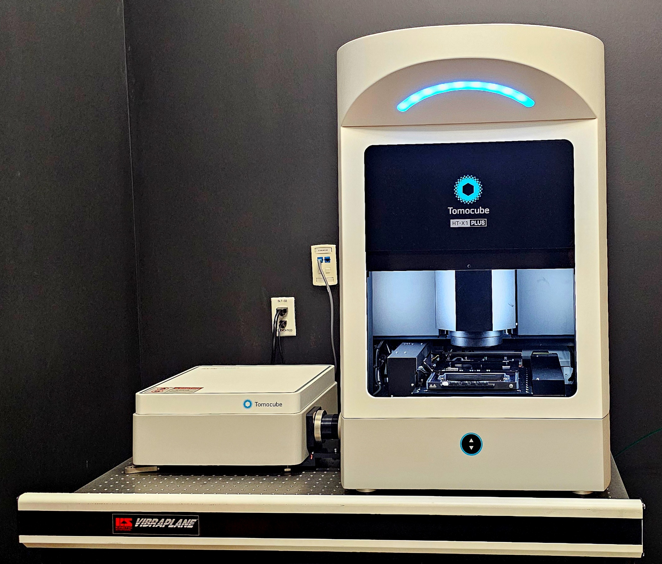
Tomocube HT-X1 Plus
Holotomography · Inverted
Label-free 3D holotomography system with fluorescence imaging for live cells, organoids, and tissue.
Manufacturer: Tomocube
Location: NL5.120S
Internal rate: $30/hour
Standard internal rate
Key capabilities: Holotomography: label-free imaging; Widefield fluorescence; 3D refractive-index mapping; Focus stabilization
Technical details ▾
| Imaging modalities | Holotomography; Widefield fluorescence; Brightfield |
|---|---|
| Specimen types | Live cells; Organoids |
| Sample formats | Slides; 35 mm dishes; 50 mm dishes; Multi-well chambers; 6/12/28 microtiter plates. Not compatible with standard 96-well plates. Glass bottom #1.5 required. |
| Excitation | LEDs: 405; 488; 561; 640 |
| Detectors | 20 MP CMOS |
| Cameras | sCMOS |
| Transmitted light | Label-free refractive-index imaging |
| Environmental control | Live-cell: Yes; temperature: Yes; CO₂: Yes; humidity: Yes |
| Special capabilities | 3D refractive-index mapping; Label-free imaging; Focus stabilization |
| Objectives | 40x/0.95 air |
| Recommended applications | Label-free live-cell imaging; Organoids; Tissue; Correlative fluorescence and holotomography |

Zeiss LSM780 with 2-P
Confocal · Inverted
Inverted spectral confocal and multiphoton microscope with live-cell incubation.
Manufacturer: Zeiss
Location: NL5.120N
Internal rate: $40/hour
Half price outside internal peak hours
Key capabilities: Laser-scanning confocal; Multiphoton; Spectral imaging; SHG; FRAP; Photoablation, Photoactivation
Technical details ▾
| Imaging modalities | Laser-scanning confocal; Multiphoton; Spectral imaging; SHG |
|---|---|
| Specimen types | Immunofluorescent cell cultures; Tissue sections, Organoids; Live cells; Fixed cells |
| Sample formats | Slides; 35 mm dishes; Multi-well chambers; Multi-well plates |
| Excitation | 405; 458; 488; 514; 561; 594; 633 |
| Multiphoton range | 690–1040nm |
| Detectors | 32-channel GaAsP PMT Array; 2 Standard Multi-Alkali (MA) PMTs; 2 non-descanned detectors |
| Environmental control | Live-cell: Yes; temperature: Yes; CO₂: Yes; humidity: Yes |
| Special capabilities | Simultaneous multi-channel acquisition; Focus stabilization; Automated z-stacks; Tiling; FRAP; Photoablation; Photoactivation; Uncaging; Spectral unmixing; FRET; SHG. |
| Objectives | 10x/0.3; 20x/0.8; 25x/0.8; 40x/1.4; 63x/1.4 |
| Recommended applications | Confocal imaging; Live-cell imaging; Fixed-cell imaging; Multiphoton imaging; Deep-Tissue, Organoids imaging; FRET; Spectral unmixing; SHG |

Zeiss LSM780 upright with 2-P
Confocal · Upright
Upright confocal and multiphoton microscope configured for organ culture, deep Tissue, Organoids, and whole-animal imaging.
Manufacturer: Zeiss
Location: ND5.740
Internal rate: $40/hour
Half price outside internal peak hours
Key capabilities: Laser-scanning confocal; Multiphoton; Spectral imaging; SHG; FRAP; Deep imaging; Motorized stage
Technical details ▾
| Imaging modalities | Laser-scanning confocal; Multiphoton; Spectral imaging; SHG |
|---|---|
| Specimen types | Organ culture; Whole-animal preparations; Thick Tissue, Organoids |
| Excitation | 405; 458; 488; 514; 561; 594; 633 |
| Multiphoton range | 690–1040 nm |
| Detectors |
32-channel GaAsP PMT Array; 2 Standard Multi-Alkali (MA) PMTs; 2 non-descanned detectors |
| Environmental control | Temperature: Yes |
| Special capabilities | Deep imaging; FRAP; FRET; Photoablation; Photoactivation; Uncaging; Spectral unmixing; SHG; Simultaneous multi-channel acquisition; Automated z-stacks; Tiling |
| Objectives | 10x/0.3; 10x/0.45; 20x/0.8; 20x/1.0; 32x/0.85; 40x/1.1; 40x/1.3; 63x/1.4 |
| Recommended applications | Organ culture; Whole-animal imaging; Deep-Tissue, Organoids imaging; Multiphoton imaging; SHG |

Zeiss LSM880 Airyscan
Confocal · Inverted
Super-resolution
Inverted laser-scanning confocal microscope with 32-channel spectral detection and Airyscan super-resolution.
Manufacturer: Zeiss
Location: K1.224
Internal rate: $40/hour
Half price outside internal peak hours
Key capabilities: Laser-scanning confocal; Spectral imaging; FRAP; Photoablation; Photoactivation; Uncaging
Technical details ▾
| Imaging modalities | Laser-scanning confocal; Spectral imaging |
|---|---|
| Super-resolution | Airyscan FAST |
| Specimen types | Immunofluorescent cell cultures; Tissue sections, Organoids; Live cells; Fixed cells |
| Sample formats | Slides; 35 mm dishes; Multi-well chambers; Multi-well plates |
| Excitation | 405; 458; 488; 514; 561; 633 |
| Detectors | 32-channel GaAsP PMT Array; 2 Standard Multi-Alkali (MA) PMTs; Airyscan FAST detector |
| Environmental control | Live-cell: Yes; temperature: Yes; CO₂: Yes; humidity: Yes |
| Special capabilities | Simultaneous multi-channel acquisition; Focus stabilization; Automated z-stacks; Tiling; Airyscan; FRAP; Photoablation; Photoactivation; Uncaging; Spectral unmixing; FRET |
| Objectives | 10x/0.3; 20x/0.8; 40x/1.1; 40x/1.4; 63x/1.4 |
| Recommended applications | Confocal imaging; Super-resolution confocal imaging; Live-cell imaging; Fixed-cell imaging;Tissue, Organoid; Spectral imaging |

Zeiss LSM880 with 2-P
Confocal · Inverted
Inverted spectral confocal and multiphoton microscope with FCS, live-cell incubation, and photomanipulation tools.
Manufacturer: Zeiss
Location: NL7.134A
Internal rate: $40/hour
Half price outside internal peak hours
Key capabilities: Laser-scanning confocal; Multiphoton; Spectral imaging; SHG; FCS; FRAP
Technical details ▾
| Imaging modalities | Laser-scanning confocal; Multiphoton; Spectral imaging; SHG; FCS |
|---|---|
| Specimen types | Immunofluorescent cell cultures; Tissue sections, Organoids; Live cells; Fixed cells |
| Sample formats | Slides; 35 mm dishes; Multi-well chambers; Multi-well plates |
| Excitation | 405; 458; 488; 514; 561; 594; 633 |
| Multiphoton range | 690–1040 |
| Detectors | 32-channel GaAsP PMT spectral detector; Dual-channel non-descanned detector |
| Environmental control | Live-cell: Yes; temperature: Yes; CO₂: Yes; humidity: Yes |
| Special capabilities | FCS; FRAP; Photoablation; Photoactivation; Uncaging; Spectral unmixing; FRET; SHG; Simultaneous two-channel acquisition; Focus stabilization; Automated z-stacks; Tiling |
| Objectives | 10x/0.3; 20x/0.8; 40x/1.2; 40x/1.4; 100x/1.4 |
| Recommended applications | Confocal imaging; Live-cell imaging; Fixed-cell imaging; Tissue, Organoid; Spectral imaging; Multiphoton imaging; Deep-Tissue, FCS; FRET; Spectral unmixing; SHG |
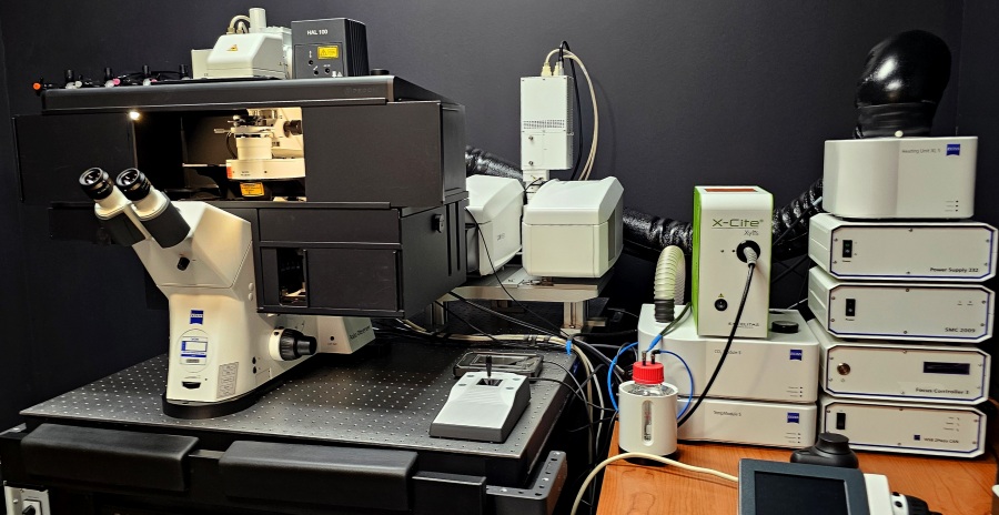
Zeiss LSM980 Airyscan 2
Confocal · Inverted
Super-resolution
Inverted spectral confocal microscope with Airyscan 2, FCS components, automated sample finding, and fast acquisition workflows.
Manufacturer: Zeiss
Location: NL5.120LA
Internal rate: $40/hour
Half price outside internal peak hours
Key capabilities: Laser-scanning confocal; Spectral imaging; FCS; Airyscan 2; FRAP
Technical details ▾
| Imaging modalities | Laser-scanning confocal; Spectral imaging; FCS |
|---|---|
| Super-resolution | Airyscan 2 |
| Specimen types | Immunofluorescent cell cultures; Tissue sections, Organoids; Live cells, Fixed cells |
| Sample formats | Slides; 35 mm dishes; Multi-well chambers; Multi-well plates |
| Excitation | 405; 445; 488; 514; 561; 594; 639; 730 |
| Detectors | 32-channel GaAsP PMT spectral detector; 2-channel MA-PMT; T-PMT 2; Airyscan 2 detector |
| Cameras | High-end epi-fluorescence camera |
| Transmitted light | Transmitted-light detector |
| Environmental control | Live-cell: Yes; temperature: Yes; CO₂: Yes; humidity: Yes |
| Special capabilities | AI sample finder; Simultaneous multi-channel acquisition; Automated z-stacks; Tiling; Airyscan 2; FCS; FRAP; FRET; Photoablation; Photoactivation; Uncaging; Spectral unmixing; infrared imaging; Focus stabilization |
| Objectives | 10x/0.3 air; 20x/0.8 air; 25x/0.8 multi-immersion; 40x/1.2 water; 40x/1.3 oil; 63x/1.4 oil |
| Recommended applications | Confocal imaging; Super-resolution confocal imaging; Live-cell imaging; Fixed-cell imaging; Tissue, Organoid; Spectral imaging; FCS; FRET; Spectral unmixing |

Zeiss SteREO Discovery V12
Stereo/dissecting · Stereo
Motorized fluorescence stereo microscope for screening and documenting whole-mount organisms, organs, Tissues, Organoids.
Manufacturer: Zeiss
Location: NL7.110M
Internal rate: $15/hour
Standard internal rate
Key capabilities: Fluorescence stereo microscopy; Brightfield; Extended-depth-of-field imaging; Automated z-stacks; Extended focus; Simultaneous GFP and transmitted-light imaging
Technical details ▾
| Imaging modalities | Fluorescence stereo microscopy; Brightfield; Extended-depth-of-field imaging |
|---|---|
| Specimen types | Whole-mount flies; Worms; Mice; Organs; Tissue, Organoids |
| Excitation | Filter sets: GFP; RFP; Cy5 |
| Detectors | Cooled monochrome camera; Color camera |
| Cameras | Zeiss Axiocam MRm; Zeiss Axiocam MRc5 |
| Transmitted light | Brightfield; Transillumination; Gooseneck illumination |
| Environmental control | Live-cell: No; temperature: No; CO₂: No; humidity: No |
| Special capabilities | Automated z-stacks; Extended focus; Simultaneous GFP and transmitted-light imaging |
| Recommended applications | Screening; Whole-mount specimens; Dissection; Documentation; Fluorescence screening |